lv filling tamponade | tamponade removal lv filling tamponade Cardiac tamponade — or pericardial tamponade — happens when the pericardium fills with fluid (usually pericardial fluid or blood). Because the fluid has nowhere to go, your heart runs out of . In 2007, the 14000 series was replaced by the new and improved 1142XX. This reference series changed the look of the Air-King. Featuring a thicker case, the new Air-King was slightly larger. It was topped with a concentric dial and sported a brand new machined Oyster bracelet.
0 · transient buckling of tamponade
1 · tamponade testing questions and answers
2 · tamponade removal
3 · tamponade pericardial effusion
4 · tamponade management chart
5 · frank tamponade arterial line placement
6 · cardiac tamponade pdf
7 · cardiac tamponade after surgery
Rolex 2000 Rolex Datejust Two-Tone 79173, 26mm, White Dial,. for $5,000 for sale from a Seller on Chrono24.

transient buckling of tamponade
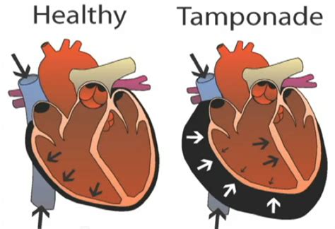
Low pressure tamponade may occur in the following situations: (1) Rapid accumulation of a pericardial effusion (e.g., acute hemorrhagic pericardial effusion). (2) Subacute pericardial effusion plus acute volume loss (e.g., hemorrhage, gastroenteritis, or fluid removal at dialysis). See moreCardiac tamponade is a clinical syndrome caused by the accumulation of fluid in the pericardial space, resulting in reduced ventricular filling and subsequent hemodynamic compromise. The.Cardiac tamponade = when pericardial effusion leads to increased pressure, impairing ventricular filling and resulting in decreased cardiac output. This is a clinical diagnosis although echo .Cardiac tamponade — or pericardial tamponade — happens when the pericardium fills with fluid (usually pericardial fluid or blood). Because the fluid has nowhere to go, your heart runs out of .
Cardiac tamponade is caused by the buildup of pericardial fluid (exudate, transudate, or blood) that can accumulate for several reasons. . In cardiac tamponade, the pericardium is too full and the LV cannot go anywhere. The inspiratory increase in RV filling results in such a bulge of the septum that the LV stroke volume is greatly diminished, with the resulting . The primary goal in the management of tamponade is to relieve the effect of the fluid or clot on cardiac pressures and restore forward flow. •. Perioperative goals are to maintain preload and systemic vascular resistance; and to avoid bradycardia and myocardial depression. Pericardial disease can present clinicians with unique challenges.
Right heart filling increases, but left ventricular (LV) filling decreases by <5% so aortic pulse pressure remains nearly constant even as peak aortic pressure falls . A hallmark of cardiac tamponade is an exaggerated inspiratory decrease in .Tamponade is characterized by elevated intrapericardial pressure (greater than 15 mm Hg), which restricts venous return and ventricular filling. As a result, the stroke volume and arterial pulse pressure fall, and the heart rate and venous pressure rise. Shock and death may result. CLINICAL FINDINGS. A. Symptoms and Signs. During tamponade, P LV is high throughout diastole, so early diastolic filling is particularly impaired, resulting in the loss of the y descent in atrial pressure and CVP and a reduction in the mitral valve E wave. Low pressure tamponade may occur in the following situations: (1) Rapid accumulation of a pericardial effusion (e.g., acute hemorrhagic pericardial effusion). (2) Subacute pericardial effusion plus acute volume loss (e.g., hemorrhage, gastroenteritis, or fluid removal at .
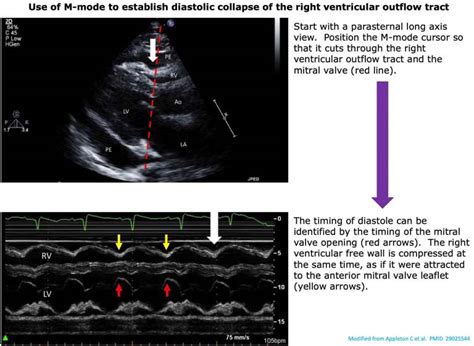
Cardiac tamponade is a clinical syndrome caused by the accumulation of fluid in the pericardial space, resulting in reduced ventricular filling and subsequent hemodynamic compromise. The.Cardiac tamponade — or pericardial tamponade — happens when the pericardium fills with fluid (usually pericardial fluid or blood). Because the fluid has nowhere to go, your heart runs out of room and can’t expand enough to fill effectively. How cardiac tamponade relates to .Cardiac tamponade = when pericardial effusion leads to increased pressure, impairing ventricular filling and resulting in decreased cardiac output. This is a clinical diagnosis although echo findings can be quite suggestive showing right sided collapse, aplethoric IVC and changes in flow across valves with respiration).
Cardiac tamponade is caused by the buildup of pericardial fluid (exudate, transudate, or blood) that can accumulate for several reasons. Hemorrhage, such as from a penetrating wound to the heart or ventricular wall rupture after an MI, can lead to a rapid increase in pericardial volume. In cardiac tamponade, the pericardium is too full and the LV cannot go anywhere. The inspiratory increase in RV filling results in such a bulge of the septum that the LV stroke volume is greatly diminished, with the resulting decrease in systolic blood pressure.
Right heart filling increases, but left ventricular (LV) filling decreases by <5% so aortic pulse pressure remains nearly constant even as peak aortic pressure falls . A hallmark of cardiac tamponade is an exaggerated inspiratory decrease in systolic blood pressure known as .
During tamponade, P LV is high throughout diastole, so early diastolic filling is particularly impaired, resulting in the loss of the y descent in atrial pressure and CVP and a reduction in the mitral valve E wave.

The true filling pressure is the myocardial transmural pressure, which is intracardiac minus pericardial pressure. 14 Rising pericardial pressure reduces and ultimately offsets this.
During spontaneous inspiration (Insp), increased filling of the RV occurs at the expense of left ventricular (LV) filling and diastolic flow across the mitral valve (E-wave) is decreased. The opposite occurs during expiration (Exp) as LV filling is enhanced. Low pressure tamponade may occur in the following situations: (1) Rapid accumulation of a pericardial effusion (e.g., acute hemorrhagic pericardial effusion). (2) Subacute pericardial effusion plus acute volume loss (e.g., hemorrhage, gastroenteritis, or fluid removal at .Cardiac tamponade is a clinical syndrome caused by the accumulation of fluid in the pericardial space, resulting in reduced ventricular filling and subsequent hemodynamic compromise. The.Cardiac tamponade — or pericardial tamponade — happens when the pericardium fills with fluid (usually pericardial fluid or blood). Because the fluid has nowhere to go, your heart runs out of room and can’t expand enough to fill effectively. How cardiac tamponade relates to .
Cardiac tamponade = when pericardial effusion leads to increased pressure, impairing ventricular filling and resulting in decreased cardiac output. This is a clinical diagnosis although echo findings can be quite suggestive showing right sided collapse, aplethoric IVC and changes in flow across valves with respiration).
tamponade testing questions and answers
Cardiac tamponade is caused by the buildup of pericardial fluid (exudate, transudate, or blood) that can accumulate for several reasons. Hemorrhage, such as from a penetrating wound to the heart or ventricular wall rupture after an MI, can lead to a rapid increase in pericardial volume. In cardiac tamponade, the pericardium is too full and the LV cannot go anywhere. The inspiratory increase in RV filling results in such a bulge of the septum that the LV stroke volume is greatly diminished, with the resulting decrease in systolic blood pressure.
Right heart filling increases, but left ventricular (LV) filling decreases by <5% so aortic pulse pressure remains nearly constant even as peak aortic pressure falls . A hallmark of cardiac tamponade is an exaggerated inspiratory decrease in systolic blood pressure known as . During tamponade, P LV is high throughout diastole, so early diastolic filling is particularly impaired, resulting in the loss of the y descent in atrial pressure and CVP and a reduction in the mitral valve E wave. The true filling pressure is the myocardial transmural pressure, which is intracardiac minus pericardial pressure. 14 Rising pericardial pressure reduces and ultimately offsets this.

new rolex yacht master rubber strap
Rolex released the first Submariner in 1953. While there is some debate about which reference was actually first, it is widely believed to have been ref. 6204. Three different Submariner references appeared within the first year of its existence, but most scholars and collectors believe the ref. 6204 was the very first based on the serial .
lv filling tamponade|tamponade removal


